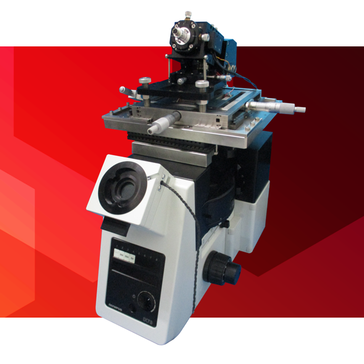
Probe-Scanning
High-Speed Atomic Force Microscope
PS-NEX
- Realize the true ‘in situ’ observation -
Atomic Force Microscope(AFM) is a powerful visualization tool. It's capable imaging real nano-scale structure in air and liquid.
But serious drawback to conventional AFM is slow scanning speed. So that the image is only still picture.
Our High-Speed AFM can observe real-time imaging as movie.
Even swaying samples in solution can be imaged clearly without blurring, since the image acquisition time is short enough.
It is unnecessary to anchor the sample tightly onto substrate, thus the adverse effect from sample preparation is minimized.
PS-NEX(Probe scan type) is evolved our High-Speed AFM SS-NEX additional features.
Observations of various samples can be realized while keeping the function of taking movies.
Simultaneous observation of fluorescence image and AFM image is also possible by integrating optical microscope.
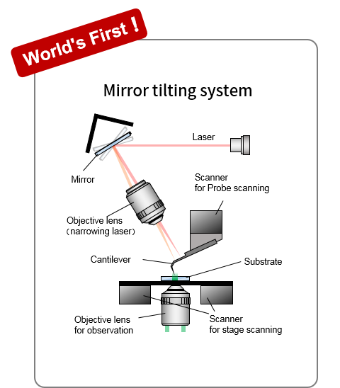
- High-speed laser tracking tilting mirror to synchronize with high-speed scanning cantilever
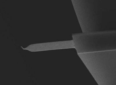
Ultra small cantilever (Olympus)
Resonance Frequency: 1500 kHz (in air), 500 kHz (in liquid)
Spring Constant: 0.1 N / m
Length: 10 μm
[ Page Top ]
| Scan system | Probe scan | High speed scanning (AFM observation) |
|---|---|---|
| Stage scan | Slow scanning (position adjustment) | |
| High speed scanning (Probe scan) | Maximum scan range | X: 4 µm, Y: 4 µm, Z: 2 µm |
| Scan speed | 150 ms / frame (6.7 frames / sec ) | |
| Stage scanning | Maximum scan range | X: 45 µm, Y: 45 µm |
| Scan speed | 60 s / frame | |
| Sample size | 36 mm x 36 mm x 3 mm (Φ 50mm) | |
| Sample stage movement range | 20 mm x 20 mm | |
| Observation environment | In liquid | |
| Probe sensing system | Optical detection system by mirror tilting (optical lever system) | |
| Observation mode | AC mode (shape image, error image, phase image) | |
| Laser | 790 nm (Infrared) | |
| Optical microscope | Standard specified product | |
| Rack, vibration isolation table | Adapted to the installation environment | |
-
Note1: The scan range of the scanner is a typical value.
-
Note2: Scan speed is determined for each scanner under specific conditions, and it does not guarantee the scan speed at the maximum scan range.
-
Note3: Regarding the options, taking into account on the user needs, the required set up will be arranged by visiting any time.
[ Page Top ]
| Fluorescence microscope |
Please enquire about selecting the different models |
|---|---|
|
Total internal reflection fluorescence microscope |
Please enquire about selecting the different models |
| Sample stage | Please contact us for your requirements |
- Note: Regarding the options, taking into account on the user needs, the required set up will be arranged by visiting any time.
[ Page Top ]