  |
|
|
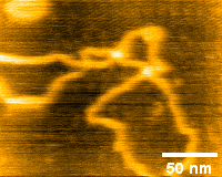
Plasmid DNA (using High-Speed Module) This is an image of plasmid DNA soft-landed on mica.
|
|
 |
 |
|
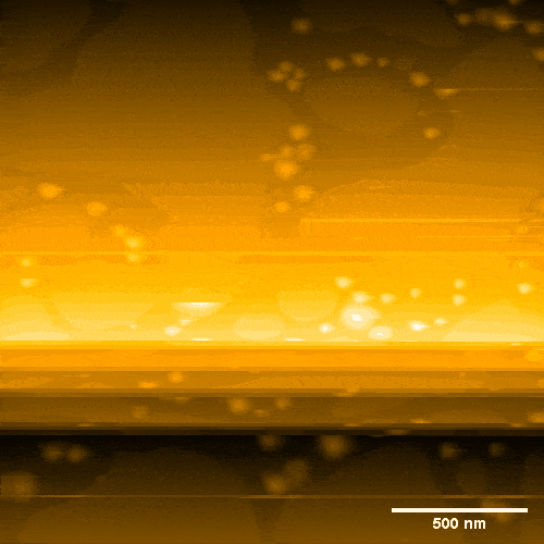
Membrane fusion of POPC This is an observed image of POPC vesicles drifting in solution.The image shows the fusion of spherical vesicles and POPC membranes by increasing the tactile pressure. |
Randomly colliding IgG This is the collisional motion of IgG molecules in a molecular crowding environment.It is possible to capture molecules in a "live" state, which is impossible to observe using conventional biochemical methods. |
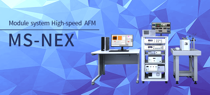
Module System High-speed AFM
The High-Speed Atomic Force Microscope (AFM) is a microscope that allows you to visualize a nanoscale sample in air or solution condition with a high-resolution movie.
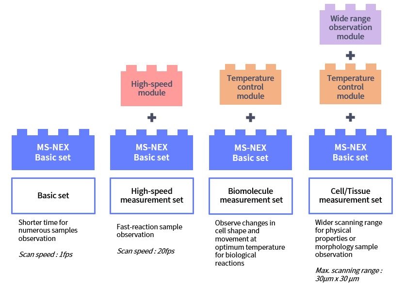
MS-NEX is based on the SS-NEX (Sample Scan type High-speed AFM) model, but with a much simpler and easier handling. It features a module system which allows you to select only the functions you need.
Various types of modules are available to be selected to fit your research and budgets.
*The High-Speed AFM was developed by Prof. Ando (Kanazawa Univ.) and commercialized by RIBM.
1. Flexible selection of various functions
MS-NEX which uses a module system make it easier to choose various functions modules that can be customized to fit your needs.
The modules can be added now, or later when needed. New developed function modules will also be in the lineup in the future.
Example of module selection
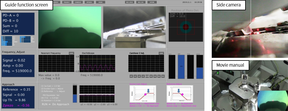
2. Guide system
MS-NEX incorporates a guide system. It is feasible even for a beginner to operate the MS-NEX. The guide system will assist from sample preparation to sample observation.
3. [Under development] Scanning speed of up to 50 fps
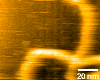
We are working on improving the High-Speed Module to achieve even higher speed.We have been able to observe plasmid DNA at a scanning speed of 50 fps.
DNA is an easy sample to observe at high speed because it is small in height and relatively fragile.
Currently, we are working on improvements to achieve high-speed observation of protein molecules, which are taller and more fragile than DNA.
Plasmid DNA at 50 fps scanning speed
Scan Speed: 20 ms / frame (50 frame / sec)
Scan Range: 100 nm × 100 nm
Image Resolution: 100 px × 100 px
[ Page Top ]
| Scan Speed |
1 s / frame (1 frame / sec) |
|---|---|
| Scan Range | XY: 4 µm × 4 µm, Z: 0.7 µm |
| Image Resolution | XY: 500 px × 500 px |
|
Observation Environment |
In air / In liquid. |
|
Observation Solution Volume |
150 μL |
| Sample Size | Φ1.5 mm |
|
Cantilever amplitude detection method |
Sample and hold method |
|
Probe Sensing Method |
Optical lever method |
| Scanning Method | Sample scan method |
| Control System | PID control, Dynamic PID control |
| Measurement Mode | AC mode (Topography image, Error image, Phase image) |
|
Software |
|
-
Note1:
Standard equipment specification includes standard scanner. -
Note2:
The maximum scanning range for each module is a typical value.
Scan speed is a value under the conditions determined for each module, and does not guarantee the scanning speed in the maximum scanning range.
[ Page Top ]
| High-speed module |
Suitable for high-speed observation such as enzyme reactions and protein structure changes.
|
|---|---|
|
Wide range observation module |
Suitable for relatively large samples with a high scanning rate.
|
|
Liquid perfusion module |
Possible to change the observation solution during AFM observation. |
|
Temperature control module |
Possible to increase the observation solution temperature from room temperature up to 40℃. |
| Illumination module |
UV, visible and other excitation light is possible to be used. |
|
Force measurement module |
Pinpoint force measurement: at a specified Position within the imaged area. Force mapping measurement: Mechanical property distribution at measurement points in a grid. |
|
OTI module |
The new scanning method enables higher speed than conventional methods. The newly developed OTI (Only Trace Imaging) method significantly reduces sample destruction. Even fragile samples can be imaged approximately 1.6 times faster. |
-
Note3:
The maximum scanning range for each module is a typical value.
Scan speed is a value under the conditions determined for each module, and does not guarantee the scanning speed in the maximum scanning range.
[ Page Top ]